STABLE™ Protocol (Soft Tissue And Bone Level Esthetics)
Conventional Implants (Flap Approach) 10-Point Clinical Checklist

Every implant surgery is a conversation between prosthesis, bone and soft tissue but too often, we only listen to one side.
The STABLE™ Protocol (Soft Tissue And Bone Level Esthetics) was born out of the need for repeatable, biologically grounded outcomes-where grafting, implant depth, soft tissue, and prosthetics align under one biologic language.
This 10-point checklist distills years of evidence-based refinement into a clear clinical framework you can follow from incision to insertion-built around one philosophy:
If the biology is stable, the esthetics follow.
In this first part, we focus on Conventional (Flap) Implants- the foundation of the protocol.
Next week, I’ll publish Part II: Immediate (Flapless) Implants- saimilar DNA, faster healing.
If you find it helpful, share with your friends
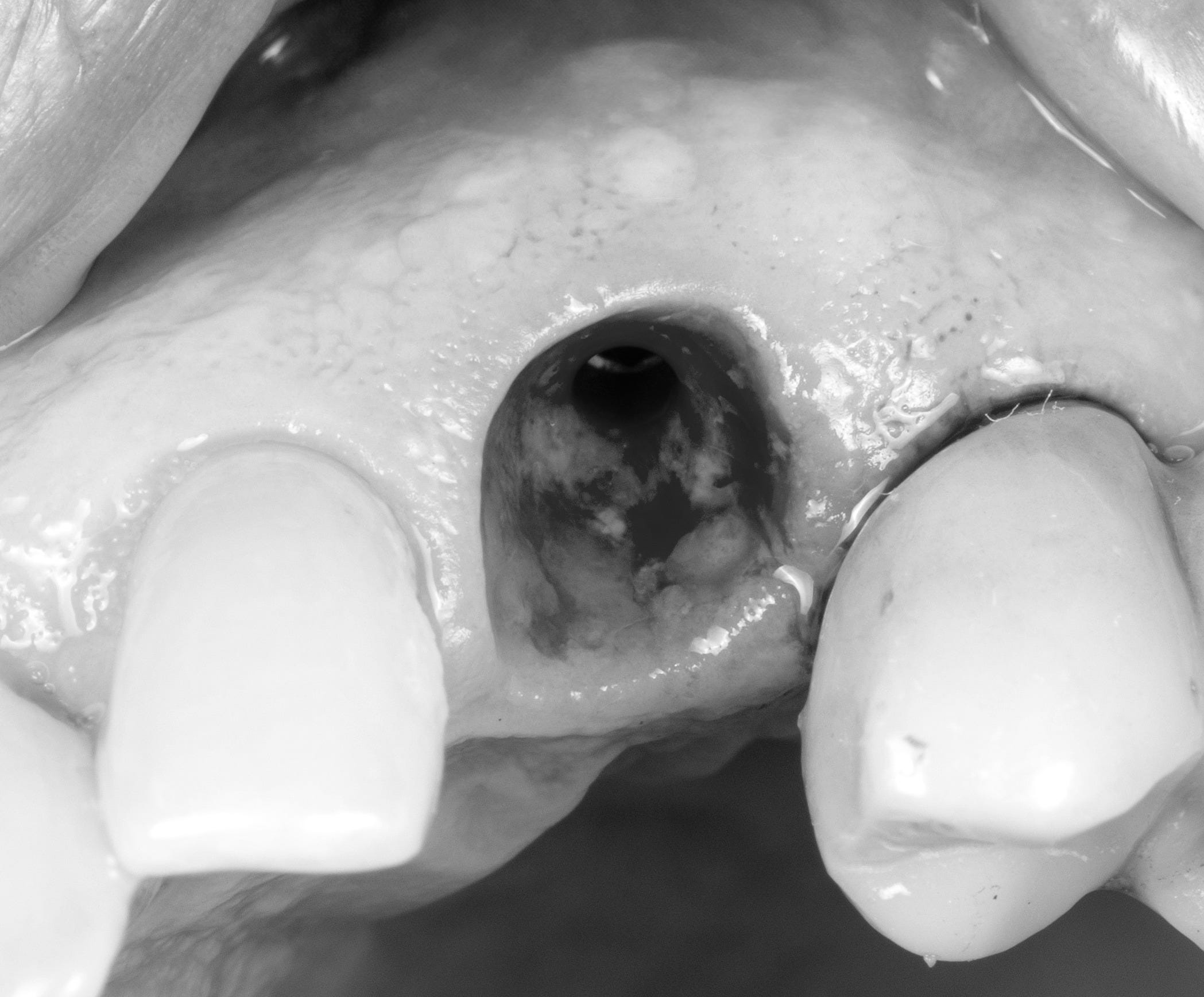
.
1️ Antibiotic Prophylaxis
Amoxicillin 2 g PO 30–60 min pre-op.
If penicillin-allergic: Azithromycin 500 mg PO 30–60 min pre-op.
→ Pre-operative prophylaxis lowers early bacterial contamination and infection risk (1).
2️ Flap Design and Asepsis
Design a full-thickness, minimally invasive flap that preserves papillae blood supply and keratinized tissue.
Reflect only as much as required for visualization; avoid extensive periosteal stripping to maintain vascular supply.
Irrigate with 1 % povidone-iodine (PVP-I) for 60 s followed by saline.
→ PVP-I reduces bacterial load and promotes fibroblast adhesion and wound healing (2).
3️ Osteotomy and 3-D Position
Prepare osteotomy following the prosthetic plan and D-I-B-A / EBC principles (3).
Position the implant platform 3–4 mm apical to the planned FGM to allow biologic-width development.
→ Correct 3-D depth and angulation determine the final soft-tissue stability (3).
Read below 4
4️ 3D space
Maintain 2mm buccal space, 1.5mm mesio-distal between teeth and 3mm inter-implant distance
If buccal plate thickness >1.5 mm → graft with fine-particle allograft (FDBA / DFDBA) and xenograft (30-70% mixture) for contour to prevent ridge resorption (4, 5).
Cover with a resorbable collagen membrane
if a cortical dehiscence is present, graft accordingly- use the STABLE grafting technique:
Inner layer (against implant or bone wall):
→ Autogenous/ allograft (if auto not possible from site near u) bone- for direct osteogenesis and rapid remodeling.
Middle layer:
→ Mixture (70:30) of autograft ± allograft + xenograft osteoconductive scaffold for volume stability.
Outer layer:
→ Membrane system (either:
Double resorbable membranes for contained defects, or
Non-resorbable (ePTFE or Ti-reinforced) + resorbable over it for dehiscence-type defects).
Stabilized via periosteal sutures or fixation tacks or both.
Build 30 % more volume than required to compensate shrinkage- periosteal release and get primary closure. Start with non-resorbable horizontal mattress and then single interrupted. Mobilise lip, cheek or tongue to see if flap moves- to avoid flap dehiscence.
5️ Primary Stability
Aim for ≥ 35 N cm insertion torque for one-stage healing.
Ensure implant micromotion < 100 µm to maintain osseointegration (6).
6️ Soft-Tissue Enhancement
Perform CTG if mucosal thickness < 2 mm or to enhance mid-facial volume (7).
Maintain ≥ 2 mm keratinized tissue buccally; perform FGG if inadequate (8).
→ Both mucosal thickness (9) and keratinized width (8) correlate with reduced crestal bone loss and inflammation.
7️ Healing Abutment and Socket Conditioning
Select the following diameter:
6-6.5mm healing abutment for molar
4.5mm for premolars, canine/ incisors
4mm or less for lateral and mandibular incisors
Select the following length:
4-5 mm to be just above mucosal margin.
Gingival height based on depth of subcrestal placement
Place a healing abutment flush with the mucosal margin; ensure tension-free closure.
Optional: use a custom healing abutment designed per EBC contour (3).
8️ Provisionalization (Early or Delayed)-optional
After soft-tissue stabilization (≈ 8–12 weeks), deliver a provisional restoration with ideal critical and subcritical contours (10).
Minimal occlusion to avoid microstrain (11).
→ Proper provisional contour guides papilla formation and mucosal scallop (10, 11).
9️ Post-Operative Protocol
Continue antibiotics for 5 days (1)- if bone grafted/ MHx or immunocompromised.
Analgesia: combination acetaminophen + ibuprofen for optimal pain control (12).
Encourage protein intake 1.2–1.6 g/kg/day (13) and optional vitamin C 500 mg/day × 1 week (14).
Rinse twice daily with 0.12 % chlorhexidine for 10 days 24 hrs after surgery.
10 Definitive Prosthesis
Confirm osseo-integration at 12–16 weeks clinically and radiographically.
Select stock abutment 2 mm gingival height.
Use ultra-polished zirconia (no glaze) at the sulcular zone to minimize plaque adhesion (15).
🧾 Key Biologic Objectives
Maintain ≥ 4 mm soft-tissue thickness for crestal bone stability (16).
Achieve tension-free flap adaptation and secure vascular integrity.
Coordinate bone level + soft-tissue level + emergence contour for biologically stable, esthetic outcomes- the foundation of STABLE™.
Methods Appendix References
Esposito M et al. Cochrane Database Syst Rev. 2013;(7):CD004152.
Bigliardi PL et al. Dermatology. 2017;233(2-3):115-126.
Esquivel-Upshaw JF et al. Int J Prosthodont. 2021;34(4):472-480.
Botticelli D et al. Clin Oral Implants Res. 2004;15(5):556-562.
Cardaropoli G et al. J Clin Periodontol. 2005;32(8):845-852.
Trisi P et al. Clin Oral Implants Res. 2016;27(12):1483-1490.
Thoma DS et al. J Clin Periodontol. 2014;41(Suppl 15):S146-S162.
Lin GH et al. J Periodontol. 2013;84(12):1755-1767.
Linkevičius T et al. Clin Oral Implants Res. 2015;26(10):1234-1238.
Chu SJ et al. Int J Periodontics Restorative Dent. 2020;40(3):377-385.
Choquet V et al. J Prosthet Dent. 2001;85(4):435-443.
Derry CJ et al. Cochrane Database Syst Rev. 2013;(6):CD010210.
Phillips SM et al. J Appl Physiol. 2017;122(4):876-881.
Pullar JM et al. Nutrients. 2017;9(8):866.
Scarano A et al. J Clin Periodontol. 2004;31(7):620-624.
Linkevičius T et al. Clin Oral Implants Res. 2015;26(10):1234-1238.